Office Hours: Mon - Wed: 8 a.m. – 12 p.m., 1 p.m. – 4:30 p.m. | Thu - Fri: 8 a.m. – 12 p.m.

Examinations

Examinations

If reflux symptoms persist over a prolonged period or recur after discontinuation of medical therapy, a structured diagnostic evaluation is recommended.

Upper endoscopy (gastroscopy) is central to the diagnostic evaluation. This allows direct assessment of inflammatory changes, Barrett’s mucosa, and the presence of a hiatal hernia. If indicated, tissue samples (biopsies) are obtained to allow for a more detailed evaluation of the mucosa.

For a functional assessment of the junction between the esophagus and the stomach, as well as the esophagus as a whole, additional testing may be required depending on the clinical situation.

A key role may be played by 24-hour esophageal pH monitoring. Using a thin catheter, acid exposure within the esophagus is measured. Alternatively, prolonged monitoring over 48 hours can be performed using a small capsule (e.g., Bravo Capsule). This allows objective quantification of the frequency, duration, and extent of acid reflux.

If necessary, esophageal manometry is performed. This test measures esophageal motility and coordination and is particularly important prior to surgical treatment, as impaired function must be taken into account when selecting the appropriate surgical approach.

In selected cases, additional radiologic evaluation of the esophagus with contrast medium may be performed. This allows assessment of swallowing dynamics, esophageal function, and the passage of contrast into the stomach and upper small intestine.
If required, the examination may be supplemented with solid food (e.g., bread or rusks) in order to assess function under more physiologic, real-life conditions.
This is particularly helpful in cases with inconclusive findings, complex clinical situations, or after previous surgery, providing additional important diagnostic information.

These diagnostic tests enable an objective assessment of reflux disease and form the basis for an individualized treatment strategy.

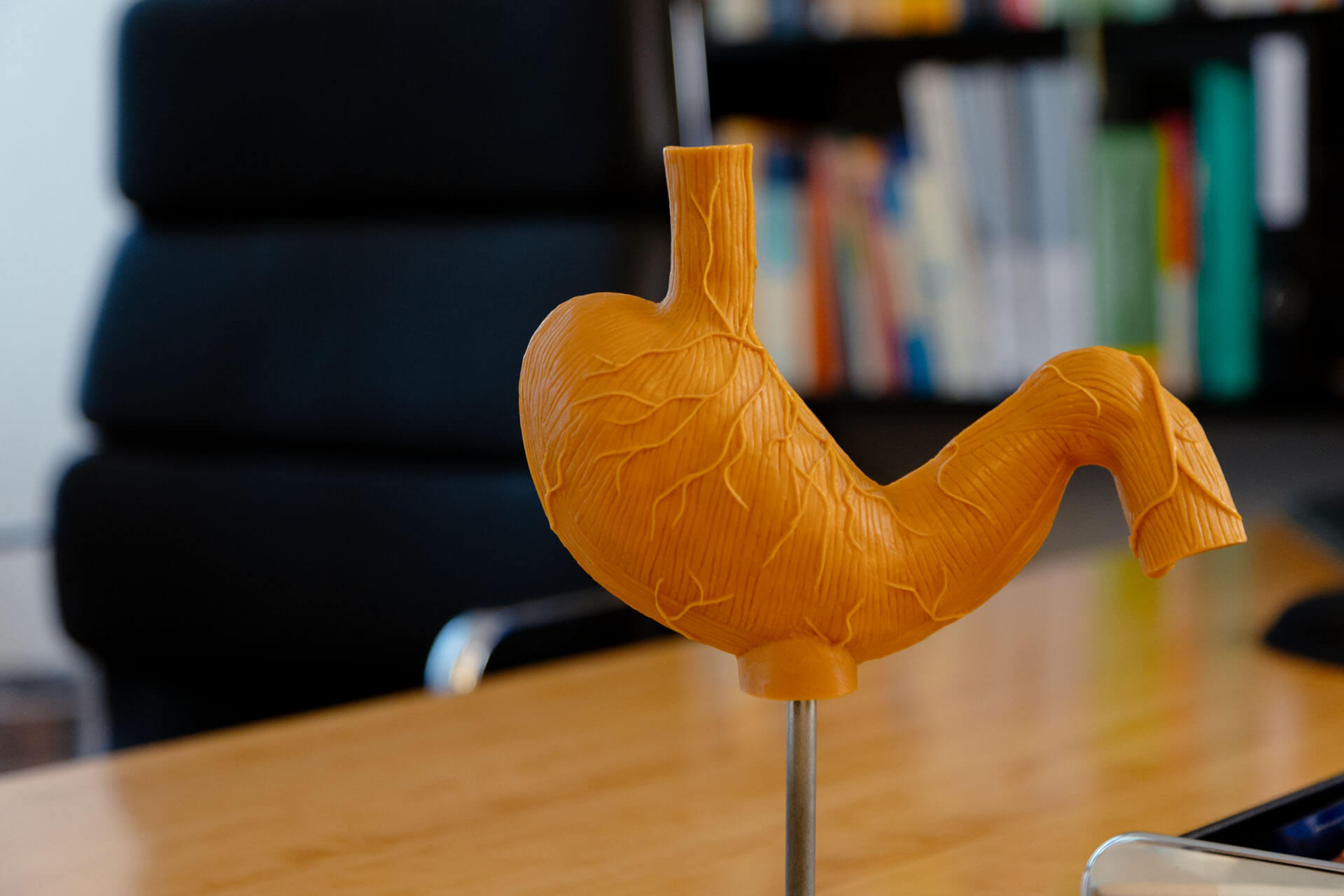
Endoskop zur Magenspiegelung

Gastroscope

Hiatal Hernias: Classification

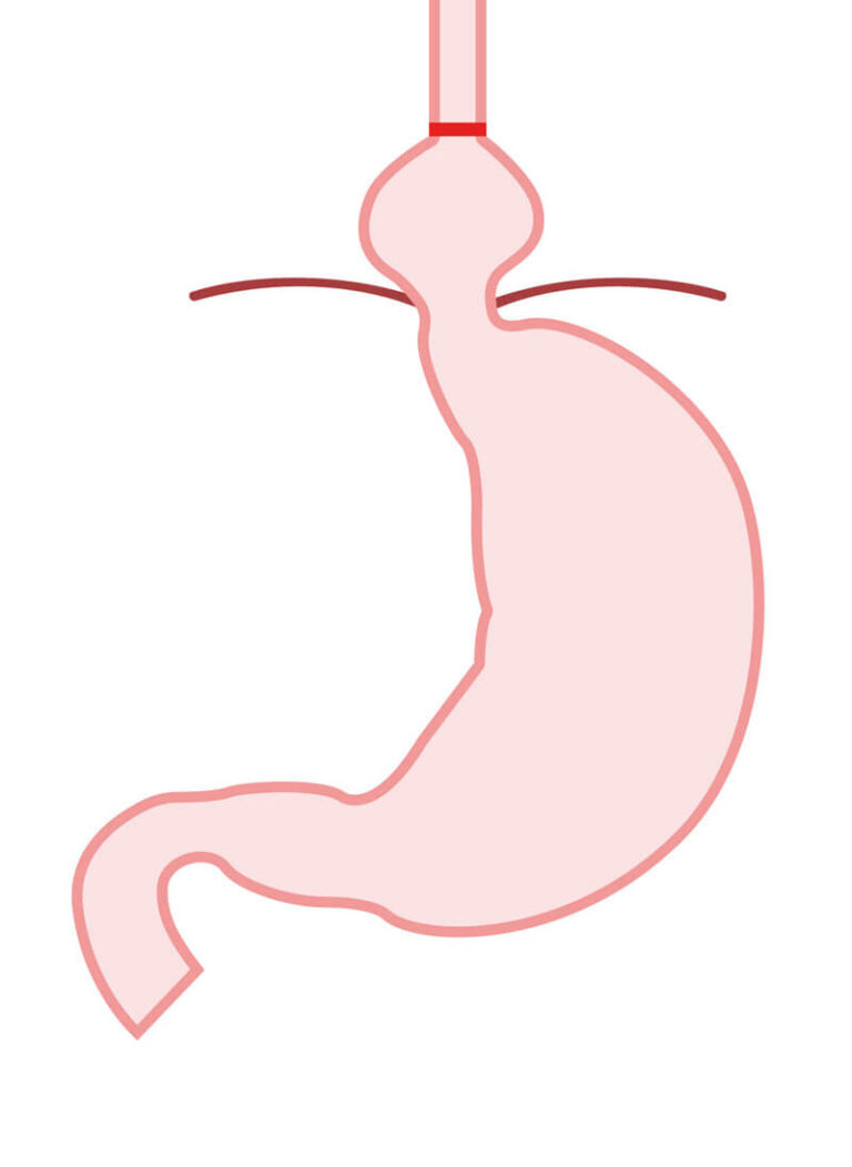
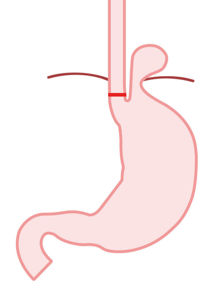
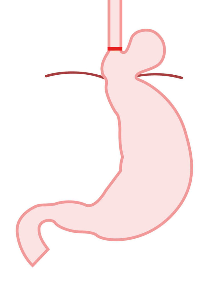
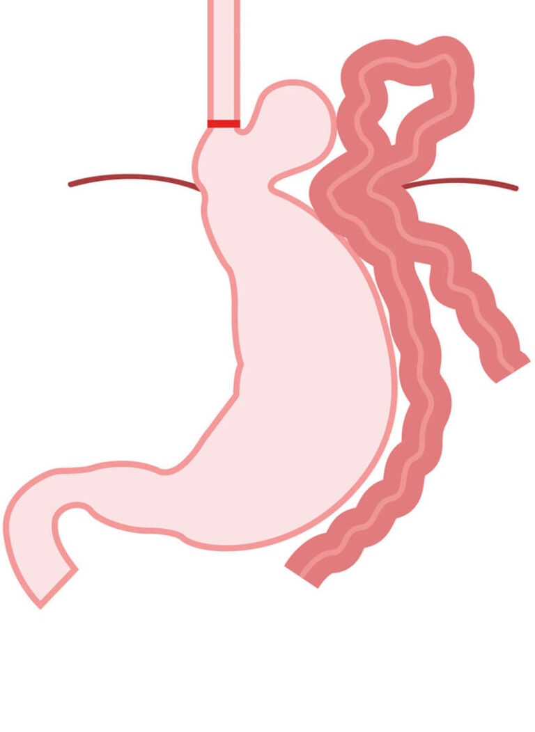
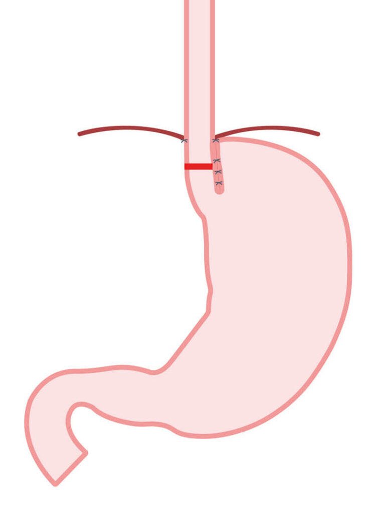
The illustrations below demonstrate the different types of hiatal hernias and their characteristic anatomical features. For comparison, the final illustration shows the anatomical appearance following successful reconstruction using our modified BICORN procedure.

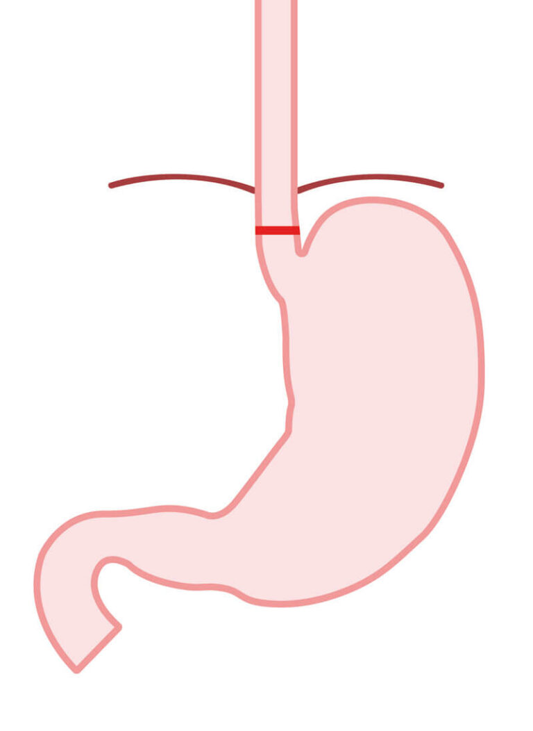
Normal Anatomy

The esophagus passes through the diaphragm into the abdominal cavity. The junction between the esophagus and the stomach is located entirely below the diaphragm.

Type I – Sliding Hiatal Hernia

The junction between the esophagus and the stomach, together with the upper part of the stomach, slides upward through the diaphragmatic opening into the chest.

Type II – Paraesophageal Hiatal Hernia

The junction between the esophagus and the stomach remains in its normal position below the diaphragm. However, part of the stomach protrudes through the diaphragmatic opening alongside the esophagus into the chest.

Type III – Mixed Hernia

Both the junction between the esophagus and the stomach and a larger portion of the stomach are located above the diaphragm. This type combines the features of both sliding and paraesophageal hiatal hernias.

Type IV – Complex Hernia

In addition to the stomach, other abdominal organs, such as the colon, small intestine, spleen, or, less commonly, other organs, herniate through the markedly enlarged esophageal hiatus into the chest. The illustration shows a large mixed hiatal hernia with part of the colon herniating through the enlarged esophageal hiatus into the chest.

Status post Modified BICORN Procedure:

The stomach is located entirely within the abdominal cavity. The junction between the esophagus and the stomach lies below the diaphragm. In contrast to normal anatomy, the angle of His is intentionally slightly accentuated to provide additional support for the body’s natural antireflux barrier.

Endoscopic Findings in Reflux Disease

Upper endoscopy (gastroscopy) is one of the most important investigations when gastroesophageal reflux disease (GERD) or a hiatal hernia is suspected. It provides valuable information about the esophagus, gastroesophageal junction, stomach, and duodenum.

It is important to understand that symptoms and objective findings do not always correlate. Some patients with large hiatal hernias experience little or no reflux symptoms, while others may suffer from severe reflux symptoms despite having no hiatal hernia or only a very small one. Likewise, the severity of reflux symptoms often does not reflect the grade of esophagitis seen.

The following images illustrate common endoscopic findings that may be encountered in patients with reflux disease.

Los Angeles Classification (LA Classification)

The severity of reflux esophagitis is usually assessed using the Los Angeles Classification (LA Classification). It is the internationally accepted standard for grading inflammatory changes of the esophagus seen during upper endoscopy.

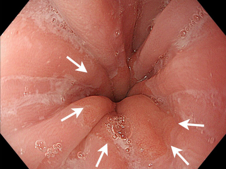
Normal Finding:

Normal endoscopic appearance without hiatal hernia, cardia insufficiency, or inflammation. The junction between the esophagus and stomach, known as the Z-line, is clearly visible. It has a slightly irregular, zigzag appearance and marks the transition between esophageal and gastric mucosa. No inflammatory changes or other abnormalities are present.

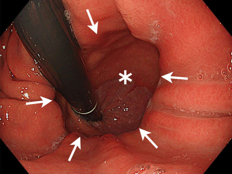
Hiatal Hernia:

Retroflexed endoscopic view obtained during upper endoscopy. The gastroscope (black) passes through the esophagus into the hiatal hernia and then into the stomach. By bending the tip of the endoscope back almost 180 degrees, the gastroesophageal junction can be viewed from below.
A moderate to large hiatal hernia (star marking) is visible, along with a markedly enlarged diaphragmatic hiatus (arrow markings).
Reflux Esophagitis According to the LA Classification

Grade A:

Normal endoscopic appearance without hiatal hernia, cardia insufficiency, or inflammation. The junction between the esophagus and stomach, known as the Z-line, is clearly visible. It has a slightly irregular, zigzag appearance and marks the transition between esophageal and gastric mucosa. No inflammatory changes or other abnormalities are present.

Grade B:

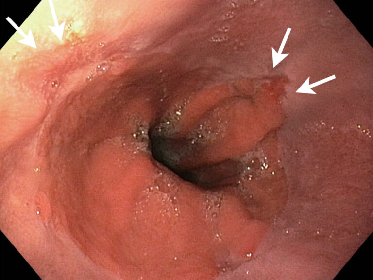
Multiple mucosal breaks are visible in the lower esophagus.

Grade C:

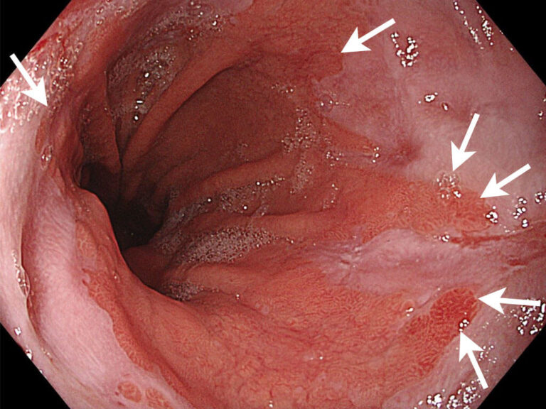
Severe inflammation and damage of the esophageal mucosa in the lower portion of the esophagus. The mucosal breaks extend over larger areas and may merge with one another. This represents an advanced stage of reflux esophagitis.

Grade D:

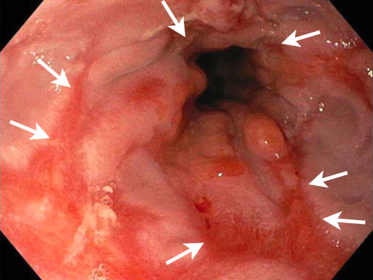
Extensive inflammation and injury of the esophageal mucosa caused by longstanding acid reflux. In the affected segment, inflammatory changes involve nearly the entire circumference of the esophagus. This represents the most severe stage of reflux esophagitis according to the Los Angeles classification.

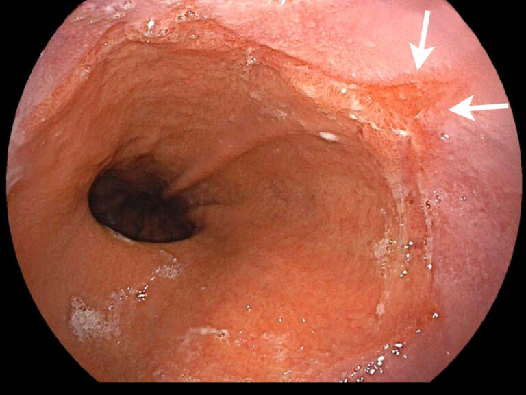
Barrett’s Esophagus

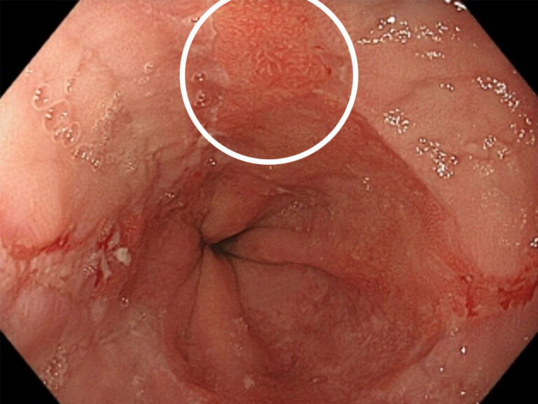
Salmon-colored mucosal tissue in the lower esophagus that is clearly distinct from the surrounding normal esophageal mucosa. This finding is characteristic of Barrett’s esophagus.

Barrett’s Adenocarcinoma

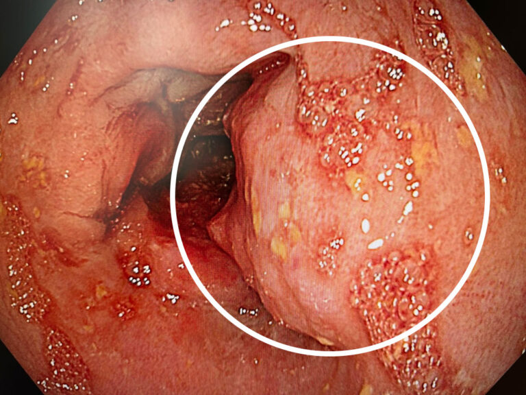
Cancer arises in an area of Barrett’s esophagus. The irregular nodular mucosal lesion protrudes into the lumen of the esophagus and causes partial narrowing of the passage.

Do you have any questions? Contact us.

Mischa Feigel, MD
Board-certified surgeon (FMH)
Specialist in visceral surgery

Florastrasse 50
CH-8008 Zurich
Google Maps directions

Office Hours

Mon - Wed:
8 a.m. – 12 p.m.
1 p.m. – 4:30 p.m.

Thu - Fri:
8 a.m. – 12 p.m.